Is Nuclear Membrane In Plant And Animal Cells
This page content
1. Components
ii. Function
3. Mitosis
4. Nuclear position
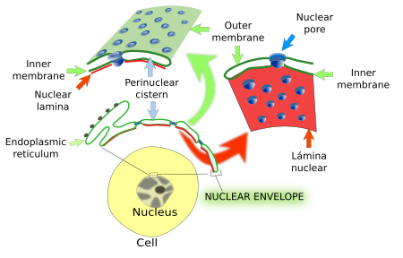
In the late Xix century, a barrier limiting the nucleus was suggested, which was later confirmed by electron microscopy (Figure 1). The nuclear envelope is equanimous of ii membranes performing a number of functions: a) it is a concrete barrier that separates nucleoplasm (chromatin and the rest of the molecular content of the nucleus) from cytoplasm; b) information technology controls the communication betwixt both, that is the motion of molecules betwixt the nucleoplasm and cytoplasm; c) it is in accuse of the nuclear morphology; d) it contributes to the inner arrangement of the nucleus providing the anchoring points where chromatin is fastened. Nuclear envelope is connected to the cytoskeleton, microtubules and actin filaments, to set up the position of the nucleus in the jail cell.

one. Components

The nuclear envelope is composed of ii membranes, outer and inner membranes, and an intermembrane space (25-40 nm in height) between them, too known equally perinuclear space. All together, they grade the and then-chosen perinuclear cisternae (Figure ii). The outer membrane is continuous with the membrane of the endoplasmic reticulum. This membrane continuity communicates the lumen of endoplasmic reticulum and the lumen of perinuclear cisternae. Thus, the nuclear envelope may work as a calcium-storing compartment, along with the endoplasmic reticulum. Attached ribosomes to the outer membrane are observed. The inner membrane has a singled-out molecular limerick. For case, there are transmembrane proteins linked to chromatin and to the nuclear lamina, another component of the nuclear envelope (see beneath). The inner and outer membranes are continuous at the nuclear pore complexes insertion places. How is it possible that outer and inner membranes accept different molecular composition? A selective retaining molecular machinery in the inner membrane has been suggested. Transmembrane proteins are synthesized in the outer membrane of the nuclear envelope or in the rough endoplasmic reticulum, and arrive to the inner membrane past lateral improvidence (cheers to the membrane continuity), just those that become linked to chromatin or to the nuclear lamina are retained in the inner membrane of the nuclear envelope.
Idue north animal cells, the nuclear lamina is a molecular scaffold located between the inner membrane and chromatin. In mammals, nuclear lamina is around 20 - 25 nm thick. The main components of nuclear lamina are proteins known as laminas, with ii isoforms: type A (A and C laminas, which are the result of the culling splicing of the same mRNA, i.due east., the same gen) and type B (B1 and B2/B3 laminas). All of them are members of the intermediate filaments family. They are organized in a net lining the inner surface of the inner membrane of the nuclear envelope, linking the inner membrane and the chromatin. The attachment between the inner membrane and the nuclear lamina is mediated by at least 20 different types of proteins inserted in the inner membrane.
Nuclear lamina performs multiple functions. It contributes to proceed the organization of the nuclear envelope, and therefore the shape and size of the nucleus. The nuclear morphology changes when the expression of proteins of the nuclear lamina is altered, which can be observed during embryo development, jail cell differentiation and some prison cell pathologies. Nuclear lamina is also a place for anchoring the nucleus to cytoskeleton, allowing to place the nucleus in a precise location within the cell, also every bit moving the nucleus from one place to another. This connection is mediated by proteins inserted in the membranes of the nuclear envelope. The spatial distribution of nuclear pore complexes in the nuclear envelope is also influenced by the nuclear lamina. Some other office of nuclear lamina is to provide physical support for chromatin, which affects factor expression. For case, chromatin anchored to nuclear lamina is not usually transcribed. These chromatin anchored regions are different depending on the cell blazon and differentiation state of the prison cell. It is suggested that nuclear lamina-chromatin interactions are regulatory elements of gene expression. During mitosis, the nuclear envelope should be disassembled and assembled again. This process is mediated by enzymatic action (phosphorylation) over the laminas that causes breakdown of nuclear lamina, and then that microtubules can contact with chromosomes. Pathological alterations of laminas effect in the then-chosen laminopathies causing nuclear disorganization, weaker nuclear envelope structure, and eventually prison cell death.
N uclear pores are inserted in the nuclear envelope. They are in charge of the trafficking between the cytoplasm and nucleoplasm (see next page).
two. Function
Since it involves quite amount of resources, why do eukaryotic cells need to split the DNA from cytoplasm? Some of the reasons are the following:
a) G ene stability. Confining the genome inside a compartment helps to maintain the stability of genes, which is higher than in prokaryotes; it should keep in mind that at that place is a huge amount of Deoxyribonucleic acid.
b) G ene regulation. Separation of the genome from the cytoplasm allows gene regulation at a level that prokaryotes will never reach. For example, it prevents or allows the access of transcription factors to DNA. Transcription factors are proteins synthesized in the cytoplasm that regulate gene expression. They must cross the nuclear envelope to work on the DNA. The molecular mechanisms that let a transcription cistron to enter the nucleus is commonly the issue of a chain of molecules, which may starting time with the activation of a receptor located in the plasma membrane. If some step of this concatenation is stopped, the factor will non be expressed.
c) Eukaryotic genes incorporate exons and introns, pregnant that a maturation process (cutting and splicing) of the main mRNA is needed. It is dangerous to translate an unprocessed mRNA because information technology will produce malformed proteins, which may even cause pathologies. This mRNA processing takes identify in the nucleoplasm and only mature mRNA is allowed to cross the nuclear envelope.
d) Transcription and translation take place in separate compartments (nucleoplasm and cytosol, respectively) and provide an additional tool for regulating the flux of information from DNA to proteins. In this way, the transcription of a gene into mRNA does not mean immediate translation. For case, preventing a particular mRNA to cross the nuclear envelope means that the prison cell will not get the protein correct abroad. However, when needed, the synthesis of this protein will be very quick because the mRNA was already synthesized and to cross the nuclear envelope is the only remaining step for the mRNA to be translated in the ribosomes.
3. During mitosis
Inorthward nigh eukaryotic cells, the nuclear envelope breaks in little vesicles during the mitotic prophase. It is known as open mitosis because cytosolic microtubules can gain access and brand contact with chromosomes. Once chromosomes are segregated, nuclear envelope is assembled again during telophase from the membranes of the endoplasmic reticulum to course the nuclei of the two new cells (Effigy 3). In yeasts, yet, the integrity of the nuclear envelope is maintained and new nuclei are formed by strangulation, as it occurs during cytokinesis. This is considering yeasts are able to build a mitotic spindle within the nucleus. This known as closed mitosis.

four. Nuclear position
The nucleus can be found in different places of the cytoplasm depending on the jail cell type, and on the prison cell action and physiology. Sometimes the nucleus is passively displaced by other components of the cell like large lipid droplets of adipocytes and myofibrils in skeletal musculus cells, both jail cell types having the nucleus shut to the plasma membrane. In general, the nucleus is actively placed in a specific region of the cytoplasm by the interaction of the cytoskeleton with the nuclear envelope, largely by actin filaments and microtubules, simply intermediate filaments tin can participate as well. In animal cells, the nuclear envelope may be connected with the centrosome, which drags the nucleus when is moved forth the microtubules. Microtubules tin can likewise contact directly with the nuclear envelope. Movement is the result of the activity of motor proteins associated to the cytoskeleton, although short distant movements may be a consequence of the polymerization and depolymerization of cytoskeletal filaments. Proteins located in the membranes of the nuclear envelope are intermediaries between cytoskeleton and the nuclear lamina. Speeds ranging from 0.one and one µm/min have been recorded. The faster movements, 10 µm/min, are those of the pronuclei of zygotes afterward fertilization.
There are several protein complexes linking the nuclear lamina to the cytoskeleton. They include transmembrane proteins in the outer membrane, KASH proteins (nesprins in mammals), and in the inner membrane, Dominicus proteins, of the nuclear envelope. There may be more than proteins involved in making these cytoskeleton-nuclear envelope bridges (Effigy 4).

In mammals, there are v genes coding for KASH proteins, some of them showing mRNA alternative splicing that tin generate quite long poly peptide isoforms exceeding 800 kDa. KASH proteins accept very long molecular processes that extend into the cytosol and contact the cytoskeleton. SUN proteins are linked to KASH proteins in the perinuclear space (betwixt the two membranes of the nuclear envelope), and to laminas (components of the nuclear lamina) by their nucleoplasmic molecular domain.
Bibliography
Gundersen GG, Worman HJ. 2013. Nuclear positioning. Jail cell. 152: 1376-1389.
Guo T, Fang Y. 2014. Functional organization and dynamics of the cell nucleus. Frontiers in institute biology. 5: 378. doi: 10.3389/fpls.2014.00378
Rowat Ac, Lammerding J, Herrmann H, Aebi U. 2008. Towards and integrated agreement of the structure and mechanics of the cell nucleus. BioEssays xxx: 226-236.
Starr DA, Fridolfsson HN. 2010. Interactions between nuclei and the cytoskeleton are mediated by SUN-KASH nuclear-envelope bridges. Almanac review of cell developmental biology. 26: 421-444.
Wanke C, Kutay U. 2013. Enclosing chromatin: reassembly of the nucleus after open up mitosis. Cell 152: 1222-1225.
Wilhelmsen K, Ketema M, Truong H, Sonnenberg A. 2006. KASH-domain proteins in nuclear migration, anchorage and other processes. Journal of jail cell science 119: 5021-5029.
Source: https://mmegias.webs.uvigo.es/02-english/5-celulas/4-envuelta.php
Posted by: sherrellsiondonsen.blogspot.com

0 Response to "Is Nuclear Membrane In Plant And Animal Cells"
Post a Comment